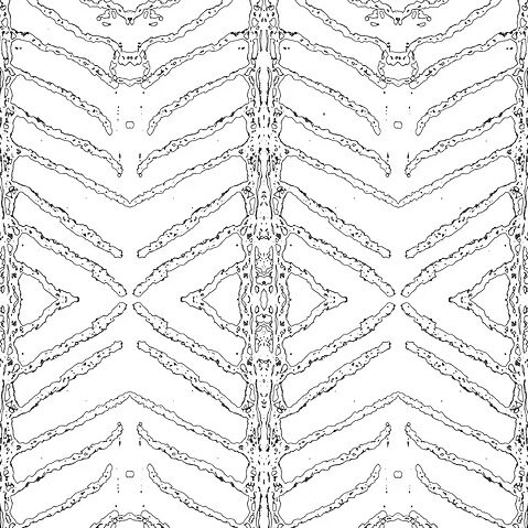
Histology is the branch of biology that studies the anatomy of biological tissues. It is through comparative studies of healthy and pathological tissues that we can arrive at a series of diagnoses. For better microscopic visualization of tissue and cell structures, most of which are colorless, they need to be stained. The most commonly used staining technique uses the hematoxylin and eosin dyes, allowing various components of the cells and the extracellular matrix to be defined quite well. In this collection, BREATHE IN WATER, we have created fun patterns based on images of histological sections of Sparus aurata (commonly known as sea bream) gills, stained with hematoxylin and eosin. The gills are respiratory organs where gas exchange takes place between the water and the blood or lymph of aquatic animals, allowing them to BREATHE IN THE WATER.
Originals *
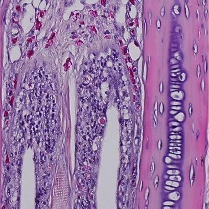
Histological section of the base of the gill filaments
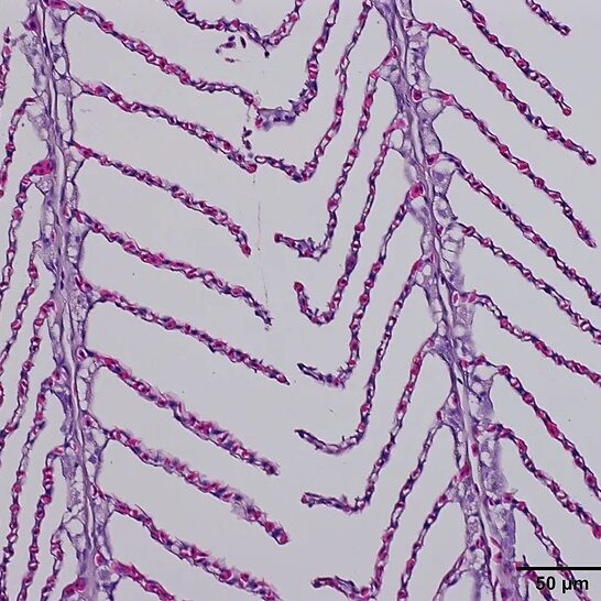
Histological section of the gill filaments
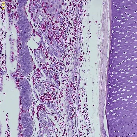
Histological section of a gill arch
*Source: Institute for Research and Innovation in Health (i3S) – Fish Immunology and Vaccinology Group
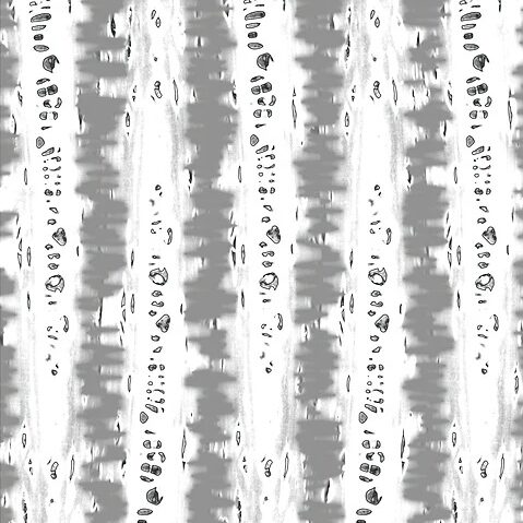
Stylized
Enjoy the collection purely as a visual voyage or as a means of understanding the science behind the images.